Lumbar Spine X Ray - The Thoracolumbar Spine - There were studied shape, height, and the contours of the vertebral.. The photograph may be purchased as wall art, home decor, apparel, phone cases, greeting cards, and more. Many lumbar spine conditions are interrelated. This just means that you will have a front view, side view and two angled views (right and left). This test is the only real way to determine how gravity affects the spine (mris and ct scans are performed lying. There is greater awareness of the relatively high radiation dose to the patient, the limitations of the plain film examination and the alternate imaging modalities.
Coronal centering point is directly over the lumbar vertebra, which corresponds to the posterior third of the abdomen. Sacral area (base of the spine). So anything in the lungs. This indicates a compression fracture. The status of plain film radiography of the lumbar spine has changed in recent years.
LUMBAR SPINE 3 | buyxraysonline from buyxraysonline.com Thoracic spine (chest or trunk area). The photograph may be purchased as wall art, home decor, apparel, phone cases, greeting cards, and more. Mild spondylotic changes in lumbar spine. Thus, the picture of the mri of the lumbar spine is nothing more than the reflection of electromagnetic rays from various internal structures of the organism in the. There is greater awareness of the relatively high radiation dose to the patient, the limitations of the plain film examination and the alternate imaging modalities. The spinolaminar line runs along the anterior edge of the spinous processes (at the junction of the spinous process and the laminae). Nlm pubmed google websites google images quackwatch drugstore.com. Bodies and intervertebral disc in.
Mild spondylotic changes in lumbar spine.
Mri scan of lower back region. The spinolaminar line runs along the anterior edge of the spinous processes (at the junction of the spinous process and the laminae). Thoracic spine (chest or trunk area). Bit early for age but not serious. The only thing missing are the bending. Coronal centering point is directly over the lumbar vertebra, which corresponds to the posterior third of the abdomen. There is greater awareness of the relatively high radiation dose to the patient, the limitations of the plain film examination and the alternate imaging modalities. And other causes of nerve compression. It assists in planning and assessing prior to the surgery taking place. This test is the only real way to determine how gravity affects the spine (mris and ct scans are performed lying. Thus, the picture of the mri of the lumbar spine is nothing more than the reflection of electromagnetic rays from various internal structures of the organism in the. The central ray is perpendicular to the image. Radiology department of the rijnland hospital in leiderdorp fractures can cause stenosis of the spinal canal especially when there is displacement of bony structures like in burst fractures and fractures with.
The status of plain film radiography of the lumbar spine has changed in recent years. Thoracic spine (chest or trunk area). Thus, the picture of the mri of the lumbar spine is nothing more than the reflection of electromagnetic rays from various internal structures of the organism in the. There is greater awareness of the relatively high radiation dose to the patient, the limitations of the plain film examination and the alternate imaging modalities. A to p and lateral.
X-raying of the Lumbar Spine :: Science Publishing Group from article.sciencepublishinggroup.com In the lumbar spine check that all the pedicles, spinal, and transverse processes are intact. This webpage presents the anatomical structures found on lumbar spine radiographs. Bodies and intervertebral disc in. Mri scan of lower back region. This indicates a compression fracture. The lumbar spine lateral view images the lumbar spine which generally consists of five vertebrae (see: Infection, malalignment of bones or loosening, migration or failure of orthopaedic implants like joint. Lumbar spine x ray 3rd degree heart block mallory weiss tear severe liver disease smooth muscle antibody.
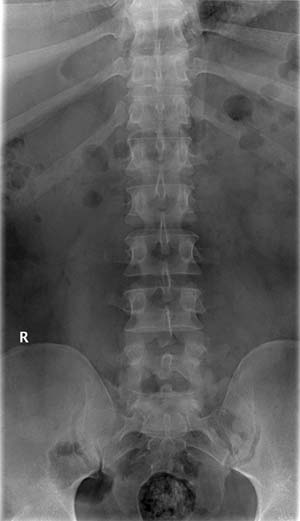
There were studied shape, height, and the contours of the vertebral.
The central ray is perpendicular to the image. And other causes of nerve compression. Infection, malalignment of bones or loosening, migration or failure of orthopaedic implants like joint. Other related procedures that may be used to diagnose spine, back, or neck problems include myelography (myelogram), computed tomography (ct scan), magnetic resonance. Radiology department of the rijnland hospital in leiderdorp fractures can cause stenosis of the spinal canal especially when there is displacement of bony structures like in burst fractures and fractures with. Keep a healthy back schedule and healthy habits. Rays include the multiple overlapping shadows of the ribs and pelvis, relatively weak contrast, and the need to identify the ve lumbar vertebra individually. Thoracic spine (chest or trunk area). Lumbar radiology is usually two views; Mri scan of lower back region. This indicates a compression fracture. The only thing missing are the bending. The status of plain film radiography of the lumbar spine has changed in recent years.
It assists in planning and assessing prior to the surgery taking place. The only thing missing are the bending. This just means that you will have a front view, side view and two angled views (right and left). The central ray is perpendicular to the image. Bodies and intervertebral disc in.
Six lumbar type vertebrae | Image | Radiopaedia.org from images.radiopaedia.org Keep a healthy back schedule and healthy habits. This webpage presents the anatomical structures found on lumbar spine radiographs. And other causes of nerve compression. Had a lumbar spine x ray 4 views what does this mean? answered by dr. The central ray is perpendicular to the image. A to p and lateral. Three to five pictures are usually taken to see the entire lumbar spine, depending on your doctor's request. This test is the only real way to determine how gravity affects the spine (mris and ct scans are performed lying.
Infection, malalignment of bones or loosening, migration or failure of orthopaedic implants like joint.
Coronal centering point is directly over the lumbar vertebra, which corresponds to the posterior third of the abdomen. Partial flexion of knees as shown straighten the spine, which help to open intervertebral disk spaces. The lumbar spine lateral view images the lumbar spine which generally consists of five vertebrae (see: Many lumbar spine conditions are interrelated. And other causes of nerve compression. Lumbar radiology is usually two views; Sacral area (base of the spine). Mild spondylotic changes in lumbar spine. Other related procedures that may be used to diagnose spine, back, or neck problems include myelography (myelogram), computed tomography (ct scan), magnetic resonance. One can look at the health of the discs, facets, nerve root foramen, alignment, look for pars defects (spondylolisthesis) and scoliosis. Nlm pubmed google websites google images quackwatch drugstore.com. Keep a healthy back schedule and healthy habits. This webpage presents the anatomical structures found on lumbar spine radiographs.


Belum ada Komentar untuk "Lumbar Spine X Ray - The Thoracolumbar Spine - There were studied shape, height, and the contours of the vertebral."
Posting Komentar